Quantification of shunt fraction using contrast ultrasound and indicator dilution in an in vitro model

Abstract
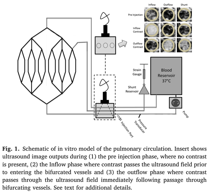
Transthoracic saline contrast echocardiography is commonly used to assess intrathoracic shunt flow in vivo. Though the technique has many advantages (safe, simple, repeatable), the measurement technique lacks specificity, and the contrast agent has limited stability. This study sought to determine if the indicator dilution modeling technique could be applied to ultrasound contrast data to quantify shunt fraction and to determine if buoyant force has a significant effect on microbubble pathway determination at a vascular bifurcation. A model of the pulmonary circuit was perfused with blood at three distinct flow rates (low, medium and high) over shunt fractions ranging from ∼2-10 %. The buoyancy effect on contrast was quantified using a simplified in vitro model of a vascular bifurcation that had an upper and lower outflow tract where saline contrast formed from carbon monoxide (CO) gas passed through the bifurcation, was collected and quantified. The indicator dilution model was found to have a mean bias of - 3.2 % for the low flow stage, - 2.6 % for the medium flow stage and - 1.4 % for the high flow stage compared to volumetric measurements, suggesting agreement increases with increasing flow rate. Investigations of the buoyant effects revealed that at lower flow rates, contrast bubbles that encounter a bifurcation will favor the upper outflow tract over the lower. However, this effect is reduced by increasing the flow rate two-fold. These data identify that application of indicator dilution theory to contrast ultrasound data and the pathway ultrasound contrast travels in a network of tubules is flow dependent.