Acute intermittent hypercapnic hypoxia and cerebral neurovascular coupling in males and females

Abstract
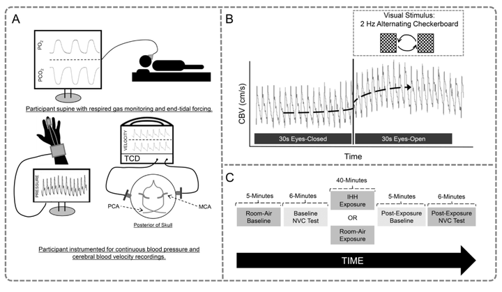
The decline in cognition observed in obstructive sleep apnea is linked to intermittent hypercapnic hypoxia (IHH), which is known to impair cerebrovascular reactivity. Whether acute IHH impairs the matching of cerebral blood flow to metabolism (i.e., neurovascular coupling, NVC) is unknown. We hypothesized that acute IHH would reduce cerebral NVC. Healthy participants (N = 17, 8 females, 9 males; age: 22 ± 3 years) had cerebral NVC measured at baseline and following 40-min of IHH at 1-min cycles with 40-s of hypercapnic hypoxia (target PETO2 = 50 mmHg, PETCO2 = +4 mmHg above baseline) and 20-s of normoxia. Cerebral NVC was quantified as the absolute and relative posterior cerebral artery blood velocity (PCAV; transcranial Doppler) and conductance (PCACVC; PCAV/mean arterial pressure [MAP]) response to a visual stimulus paradigm. Following IHH, resting PCAV was unchanged, MAP increased (+4 ± 6 mmHg, P textless 0.01) and PCACVC was reduced (−0.05 ± 0.04 cm/s/mmHg, P textless 0.01). The peak PCAV response to visual stimuli was unchanged following IHH, but the absolute and relative peak PCACVC response was increased (+0.011 ± 0.019 cm/s/mmHg, P textless 0.05 and +4.8 ± 6.1%, P textless 0.01, respectively) suggesting an increased cerebral vasodilatory response. No change occurred in the plateau cerebral NVC response following IHH. Changes in resting MAP induced by IHH did not correlate with changes in relative peak PCACVC (r2 = 0.095, P = 0.23). Cerebral NVC did not differ between sexes across all time points and was unchanged following a time-matched air-breathing control. In summary, acute IHH increases peak but not plateau cerebral NVC potentially through IHH mediated neuroplasticity.